X-ray photoelectron spectroscopy is one of the most widely used surface-sensitive electron emission spectroscopic techniques. Its development is based on the photoelectric effect, which may detect the element existing within a material or covering its surface.
X-ray photoelectron spectroscopy principle
The characteristic binding energies of core electrons in atoms keep them in their orbitals. The characteristics of adjacent atoms, orbital kinds, and atom types all influence the binding energies. The binding energies of electrons emitted from the sample surface are measured by XPS.
The mechanism of photoionization is utilized to eject electrons from surfaces. For this, the sample surface is irradiated by soft x-ray photons usually 1486 eV from an Al-anode. The photon penetrates the surface to a depth of approximately 1µm. When a photon interacts with a sample atom, the photon’s energy ionizes core electrons, causing them to be ejected from the atoms. Within the bulk of the sample, the mean free path of the ejected electrons is very small due to interaction with the surrounding atoms. But, electrons expelled from atoms in the sample’s top 5nm layer have a high chance of leaving without further interaction.
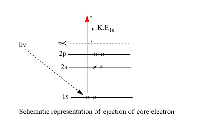
These electrons will pass into the ultrahigh vacuum(UHV) conditions within the spectrometer to ensure that electrons are not scattered by gas molecules and to limit surface contamination during the analysis. An electron energy analyzer(spectrometer) measures the kinetic energy Ek of the ejected electrons. The original binding energy Eb experienced by an electron in the sample can be calculated from Ek using the simple relationship.
Ek= hv-Eb– ϕ
where Ek= kinetic energy of the ejected electron
Eb= binding energy of the electron
hv= incident photon energy
ϕ= work function of spectrometer
The different atoms have different electron shells with different binding energies. Each element has unique binding energy (Eb) which can be used to identify the elemental composition of the sample surface. The number of emitted photoelectrons with various Ek is counted, and an XPS spectrum is generated, which facilitates the identification of all elements in all states except H and He.
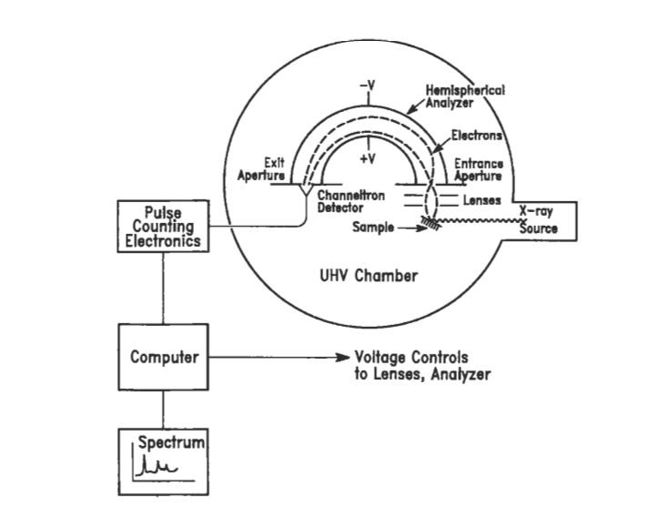
X-ray photoelectron spectroscopy instrumentation
The basic five components of the XPS instrument are listed as,
- An ultrahigh vacuum chamber(UHV) with the monochromatic soft x-ray source
- Lens system
- Energy analyzer
- Detector
- Monitor
X-ray source of XPS
Al or Mg-coated anode is struck by electrons from a high voltage around 10-15 kV, AlKα or MgKα radiation lines are produced at energies of 1486.6 eV and 1256.6 eV respectively.
Ultra-High Vacuum(UHV)
10-9– 10-10 torr or high vacuum condition is necessary to reduce the amount of noise in the XPS spectrum and to avoid any extra peaks from contaminations on the sample surface.
Electrons lens
The electron lens collects and slows the ejected photoelectrons from the sample surface before entering the energy analyzer of XPS.
Energy analyzer of XPS
It is the hemispherical sector, which consists of two concentric hemispheres with a voltage applied between them.
Detector
A multichannel display detector which is equivalent to a photographic plate allows simultaneous detection of the kinetic energy of the ejected electrons.
Monitors/Recorder
The high-performance computer is used as a recorder/monitor which controls voltage to the lense and analyzer. It also produces an XPS spectrum. The XPS spectra are generated by plotting Ek intensity vs binding energies, by computers automatically.
Application of x-ray photoelectron spectroscopy
XPS can be used for both qualitative as well as quantitative analysis.
Qualitative analysis
Elemental analysis: The electron energy levels of an atom can be divided into two types, core levels, which are bound to the nucleus, and valence levels, which are only weakly bound. The valence levels of an atom are the ones that interact with the valence levels of other atoms to form chemical bonds in molecules and compounds. Their character(valence bond character) and energy are changed by this process, becoming characteristics of the new species formed.
The core-level electrons of an atom have energies that are nearly independent of the chemical species in which the atoms are bound since they are not involved in the bonding process. Thus, the identification of Core level binding energies thus provides unique signatures of the elements. All elements in the periodic table can be identified in this manner, except for H and He which have no core levels.
Quantitative analysis
Quantitative analysis requires the measurement of relative peak intensities and the intensity of the peak depends on the atomic concentration(N) of an element present in the surface film.
Chemical shift (chemical state analysis)
Chemical shift is the difference in binding energy between the same atom in two chemically distinct states in the same molecule or in two separate compounds. It is characteristic of the nature and the number of atoms surrounding the ionized atoms of the sample. It can be used to see what kind of chemical bonding of atoms is on the sample surface.
- It concerns the chemical bonding nature, the nature of functional groups, and the oxidation states of atoms.
- An important capability of XPS is its ability to distinguish the same element in different chemical states, because of BE of an electron is different due to changes in charge density around that element by oxidation
In-depth analysis
Estimation of the in-depth distribution of elements in thin surface film is called in-depth surface analysis. It is done to know whether the thin film is homogeneous or not.
Advantages of X-ray photoelectron spectroscopy
- All samples solid, gaseous, and liquid samples can be analyzed but mostly solid samples are analyzed.
- Good quantification
- Excellent chemical state determination capabilities
- Its applicability to a wide variety of materials from biological materials to metals.
- It is a non-destructive technique.
Limitations of X-ray photoelectron spectroscopy
The main disadvantages of x-ray photoelectron spectroscopy are listed below:
- Lack of good spatial resolution i.e poor spatial resolution
- Only moderate absolute sensitivity
- Its inability to detect hydrogen and helium
- It is not suitable for trace analysis.
X-ray photoelectron spectroscopy youtube video
References
C. R. Brundle, C. K. Evans, Jr., S. Wilson and L. E. Fitzpatrick (eds), Encyclopedia of Materials Characterization: Surfaces, Interfaces, Thin Films, Butxetworch-Heinemann, Reed Publishing (USA) Inc., 199