Atomic force microscopy is a surface scanning technique that has sub-nanometer scale resolution. One disadvantage of the STM technique is that it cannot image insulating samples since a tunneling current between tip and sample is needed. The idea behind the atomic force microscope (AFM) is to measure the force(s) between the surface and the scanning tip in order to track the surface topography.
Atomic force microscopy principle
Atomic force microscope uses a sharp tip mounted on a flexible cantilever. When the tip is scanned very close to the sample surface i.e within a few Å of the sample’s surface., repulsive or attractive forces between the atoms on the tip and those on the samples are developed. The repulsive or attractive force causes the cantilever to deflect. The magnitude of the deflection depends on the tip to sample distance “d”. Several atoms on the AFM tip will interact with several atoms on the surfaces.
The displacement ( x) of the cantilever from its original position is given by
X= – f/k
Where k= spring constant of the cantilever
f = restoring force
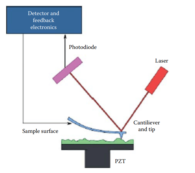
The most common method of monitoring the deflection is with an optical lever or beam-bounce detection. In this scheme, light from a laser diode is reflected from the back of the cantilever into a position-sensitive photodiode(detector). A given cantilever deflection will then correspond to a specific position of the laser beam on the positive-sensitive photodiode(detector). The back of the cantilever may be coated with gold or another metal to enhance the reflection of the laser beam into the detectors.
Modes of AFM
There are three modes of AFM. These are discussed in short below:
Contact mode AFM
In contact mode, the tip is scanned very close to the sample (distance < 10 Å) so that the repulsive force that acts between the sample and tip can be measured. Contact mode can be executed in two different ways: constant height and constant force.
In constant height mode, the position of the piezoelectric scanner is held a constant, and spatial variation of deflection of the cantilever is measured. When the cantilever gets deflected, the positions of the laser spot are reflected from the back of the cantilever.
In constant force mode, the position of the sample is varied to keep the cantilever deflections constant.
Non-constant mode
In this mode, the tip is scanned at a sample-tip distance of 10-100 Å so that attractive force is measured. In the experiment, the cantilever is allowed to oscillate at its natural frequency. When the tip comes closer to the sample (distance of 10-100 Å) the resonant frequency of the cantilever changes.
Tapping or Intermittent mode
In tapping mode, the cantilever is lowered and lifted vertically so that the tip does not stick to the surface and tip and sample damage is minimized.
Atomic force microscopy instrumentation
The instrumentation of AFM is shown in the block diagram below:
Atomic force microscopy advantages and disadvantages
Advantages/application of AFM
- Sample preparation is very easy.
- It provides accurate height information.
- It can be operated in a vacuum, under air, and in liquids.
- Biological samples or living systems can be studied by using this technique.
- It is a non-destructive technique.
- It provides a 3-D surface profile of a sample without any special surface preparation and high vacuum.
- It provides atomic-level resolution because of the force of interaction between the atoms of the tip and the sample.
Atomic force microscopy limitations
The limitations of AFM are given as:
- Tip or sample can be damaged.
- It offers a limited vertical range.
- It has a limited magnification range.
AFM youtube video
References
- C. R. Brundle, C. K. Evans, Jr., S. Wilson and L. E. Fitzpatrick (eds), Encyclopedia of Materials Characterization: Surfaces, Interfaces, Thin Films, Butxetworch-Heinemann, Reed Publishing (USA) Inc., 1992