Table of Contents
ToggleThe major difference between gram positive and gram negative bacteria are well described below:
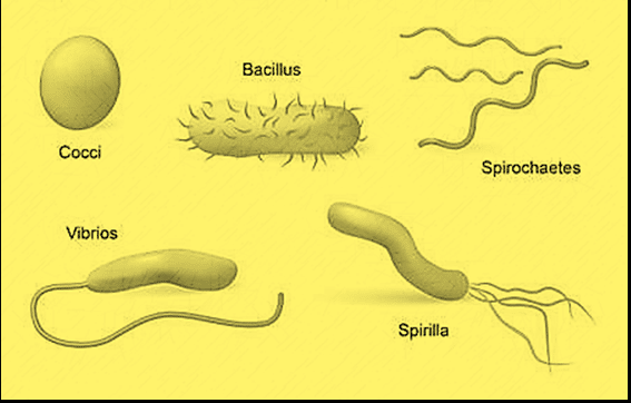
Gram positive and Gram negative bacteria are the two forms of bacteria based on the Gram’staining technique, developed by Hans Christian Grams for the identification of bacteria. As we know that bacteria are the most primitive type of organism possessing a primitive type of nucleus and are classified into various groups on the basis of staining, shape, and oxygen dependence. Bacteria are classified into five groups according to their basic shapes. They are:
- Coccus( spherical)
- Bacillus(rod)
- Spirillum (spiral)
- Comma(vibrios)
- Spirochaetes( corkscrew)

On the basis of oxygen dependence, bacteria are of two types:
- Aerobic
- Anaerobic
And on the basis of staining, they are of the following two types:
- Gram-positive
- Gram-negative
In 1884, Hans Christian Gram developed a different type of staining process based on the reaction of the cell walls with certain chemicals or dyes for the characterization and identification of bacteria. In his honor, the procedure was named “Gram stain”. Different types of reagents starting from crystal violet dye to safranine are used in this procedure.
Gram positive and gram negative bacteria
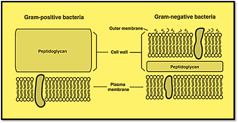
Gram-positive bacteria are the bacteria with thick cell walls (multi-layered peptidoglycan layer) that interact with gram stain retaining the crystal violet color and stain purple. Gram-negative bacteria are bacteria with thin, single-layered cell walls that cannot retain the crystal violet color due to no or very weak interaction with gram’s stain, and hence appear as light red or pink when observed under the microscope.
Mostly, they differ in their cell wall composition. The two main characteristics that influence the cell wall structure’s capacity to retain the crystal violet stain employed in the Gram staining method are the thickness of the peptidoglycan layer and the existence or absence of an outside lipid membrane. Based on these properties, gram-negative and gram-positive bacteria are identified.
Examples of gram positive and gram negative bacteria
Some examples of gram positive bacteria are Staphylococcus aureus, Streptococcus pyogenes, Bacillus subtilis, and Streptococcus pneumonia.
Pseudomonas, Klebsiella, Proteus, Salmonella, Providencia, Escherichia, Morganella, Citrobacter, etc. are some of the examples of gram-negative bacteria.
Cell wall of bacteria gram positive and negative
It is thought that gram positive bacteria have considerably larger peptidoglycans or murein, which account for the difference between the two types of bacteria. Gram positive bacteria have massive murein structures, which make up the majority (50%) of what causes the differential gram staining.
Gram negative bacteria only have a thin layer of peptidoglycans (around 20%), but they also have an extra membrane called the outer cytoplasmic membrane. Due to the increased permeability barrier caused by this, a transport mechanism across this membrane is required.

Functions of the gram-positive cell wall components
- The thick layer of murein in the gram-positive cell wall prevents osmotic lysis.
- The teichoic acid present in gram-positive bacteria helps make the cell wall stronger.
- The surface proteins in the bacterial peptidoglycans, carry out a variety of activities, some function as enzymes, and some serve as adhesions.
- The periplasmic space contains enzymes for nutrient breakdown.
Similarity between gram positive and gram negative bacteria
Both bacteria; gram positive and gram negative, have basal rings on their flagella or have flagella and capsules. These bacteria even have a cytoplasmic membrane and pilus.
The cell wall between them has a peptidoglycan layer and provides structural support to the bacteria.
Difference between gram positive and gram negative bacteria
| Characteristics | Gram-positive bacteria | Gram-negative bacteria |
| Peptidoglycan layer | Thick multi-layered | Thin single layered |
| Teichoic acid | Present | Absent |
| Periplasmic space | Absent | Present |
| Lipid and lipoprotein content | Low | High |
| Outer membrane | Absent | Present |
| Liposaccharide content | None | High |
| Dissolutionby3% KOH | Low | High |
| Spore formation | Mostly spore-forming | Mostly non- spore-forming |
| Gram stain color | Purple or blue | Red or pink |
| Antibiotic resistance | More susceptible | More resistant |
Moreover, Gram-positive bacteria produced Exotoxin as part of their growth and metabolism, whereas gram-negative bacteria produce endotoxin.
Gram stain procedure
- First, the bacteria were fixed on the slide.
- Flood the bacterial smear with staining solution i.e crystal violet and leave for 1 min
- Gently rinse with tap water or distilled water
- Flood the smear with iodine solution for 1 minute
- The smear will now appear purple.
- Decolorize the smear using alcohol and leave for 30 sec and wash with water.
- Gently flood with safranin counterstain and leave for 30 sec and rinse with tap water.
- Blot the slide dry and then observe under the microscope with oil- immersion.
Notes: Iodine and crystal violet dye were added to form a complex, which is much larger and insoluble in water. Safranin is weakly water soluble and will stain bacterial cells a light red.
Gram positive and gram negative color
Gram-positive bacteria have a distinct purple color when observed under the biological microscope due to the thick peptidoglycans layer and porous layer whereas gram-negative bacteria have a red or pink color stain.
Summary
Due to the presence of peptidoglycans layer, in their differences and structure, some bacteria are gram-positive and others are gram-negative.
Teichoic acid and lipoteichoic acids are interwoven through the peptidoglycan layer.
Gram positive are more susceptible to antibiotics and gram negative are more resistant to antibiotics. Likewise, gram positive are purple or blue in color and gram negative is pink or red in color.
For the staining of bacteria we use four different dyes they are crystal violet -iodine-alcohol- safranin.
Published by: Pratiksha Chaudhary (Chemikshya)
Gram positive and gram negative bacteria video
FAQs
1. How to identify gram positive bacteria or gram negative bacteria?
Answer: If the bacteria stay purple, they are gram-positive bacteria and if the bacteria turn pink or red, they are gram-negative bacteria.
2. Which is more harmful; gram positive or gram negative bacteria?
Answer: Gram-negative bacteria are more lethal.
3. Which bacteria are more susceptible to antibiotics?
Answer: Gram-positive bacteria are more susceptible to antibiotics.
4. How many basal rings are present in gram negative on their flagella?
Answer: Gram negative bacteria have 4 basal rings.
5. What antibiotics are used for gram negative bacteria?
Answer: Ureidopenicillins, third or fourth-generation cephalosporins, etc.
6. What colour is gram positive?
Answer: Gram positive bacteria are either purple or blue and gram negative Bacteria are either pink or red in color.
7. Which cell is stronger, gram positive or gram negative ?
Answer: Gram-positive bacteria cell is stronger.
8. Why are gram positive bacteria easier to kill?
Answer: Because of their peptidoglycans layer which is thick and absorbs antibiotics and cleaning products easily. And are more susceptible to antibiotics.


